Abdominal Blood Vessels Labeled - Solved Observe The Human Torso Model And Figures 63 7 63 9 And Chegg Com / Label the steps in the homeostatic response to high blood pressure.
Abdominal Blood Vessels Labeled - Solved Observe The Human Torso Model And Figures 63 7 63 9 And Chegg Com / Label the steps in the homeostatic response to high blood pressure.. Allows diffusion of gases and nutrients from blood into the body cells. Oxygenated blood is then returned to the left atrium of the heart by four pulmonary veins. Nerves originating from lumbar region. Although this exam has traditionally been performed with the patient. A preliminary experiment with ten ct.
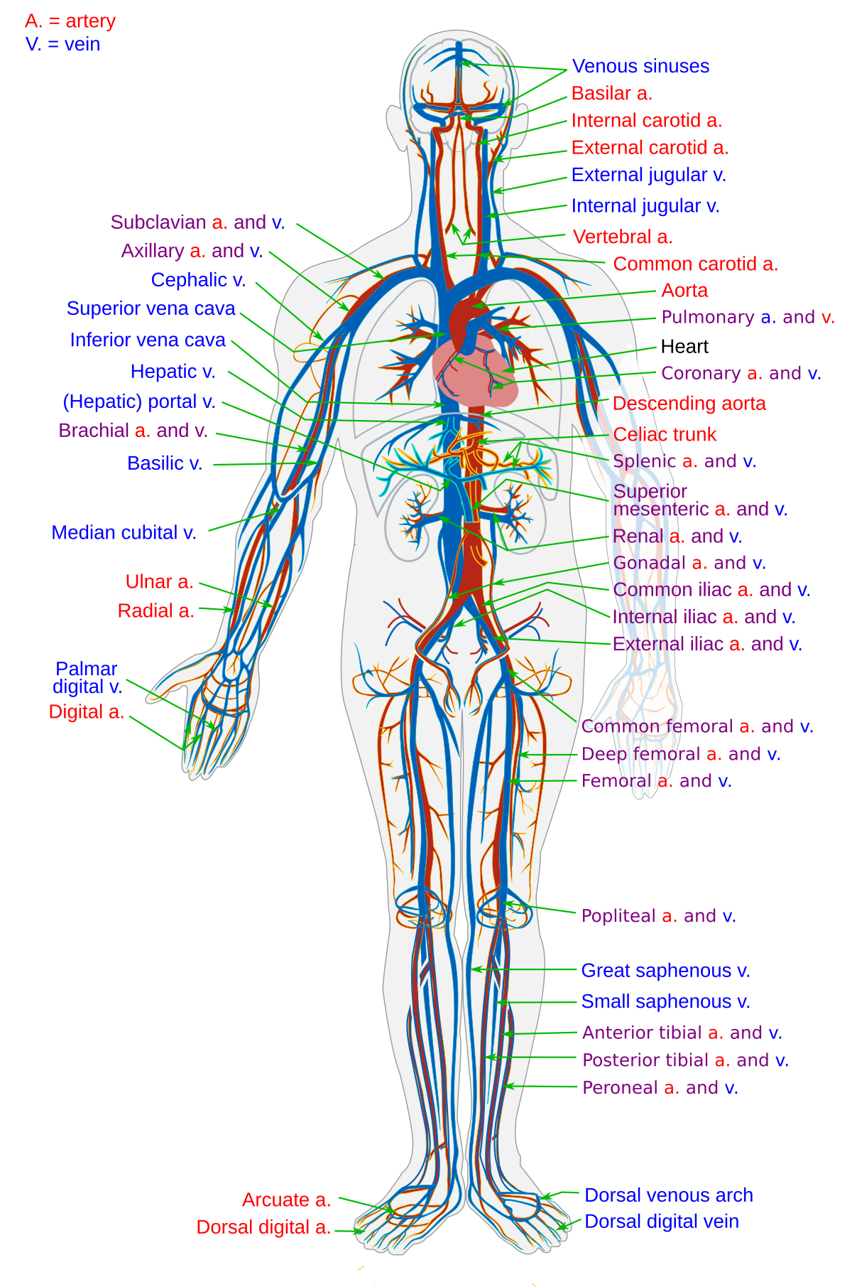
For example, new capillaries permeate the muscles of a conditioned athlete. All cells in the body need oxygen and the vital nutrients found in blood. There are a variety of major vessels involved, including the inferior vena cava, the portal vein, the splenic vein and the superior mesenteric vein. Label the veins of the upper limb. Segmentation of vessel structures in 3d volume data is of great interest for diagnosis and surgical planning.
Blood Vessel Wikipedia from upload.wikimedia.org Label the blood vessels and structures using the hints provided. Not only do blood vessels carry oxygen and nutrients, they also transport carbon dioxide and waste products away from our cells. Abdominal wall defect was prepared in 21 wistar rats. For example, new capillaries permeate the muscles of a conditioned athlete. Blood vessels are vital for the body and play a key role in diabetes helping to transport glucose and insulin. Development and function of the blood vessels: They are vital for carrying. As a medical student, i found anatomy pretty challenging.
Blood vessels (labeled) coloring page.
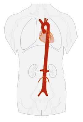
It gives off the following branches test your knowledge of the blood vessels of the abdominal aorta with the following labeling page (included in the pdf below) The descending aorta is divided into thoracic aorta and abdominal aorta by diaphragm. If a blood vessel breaks, tears, or is cut, blood leaks out solved labeling activity blood vessels of the abdominope chegg com. Researchers can now show, at molecular level, that these changes originate in vein cells. These vessels transport blood cells, nutrients, and oxygen to the tissues of the body. Posterior abdominal wall and blood vessels. They are vital for carrying. Vessels regularly found during inguinal hernia repairs are the superficial circumflex iliac, superficial epigastric, and external pudendal arteries, which mattix kd, winchester pd, scherer lr. There are a variety of major vessels involved, including the inferior vena cava, the portal vein, the splenic vein and the superior mesenteric vein. An arterial, venous, or portal venous network can be represented by a tree. Blood is oxygenated in capillaries that flow through the alveoli of the lungs. They are vital for carrying nutrients, oxygen and waste around the body. Nerves originating from lumbar region.
Blood vessels are vital for the body and play a key role in diabetes helping to transport glucose and insulin. Not only do blood vessels carry oxygen and nutrients, they also transport carbon dioxide and waste products away from our cells. Our blood vessels are not one long tube but a complex network of tubes that branch and rebranch. A preliminary experiment with ten ct. Abdominal wall defect was prepared in 21 wistar rats.
The Aorta Branches Aortic Arch Teachmeanatomy from teachmeanatomy.info All blood vessels are specifically structured to perform their function. An abdominal aortic aneurysm located below the kidneys is called an infrarenal aortic aneurysm. In abdominal surgeries, understanding blood vessel structure is critical since it is very complicated. The blood vessels make up the body's cardiovascular system. Blood is made of cells and plasma. They also take waste and carbon dioxide away from the tissues. The abdominal aorta is the largest blood vessel in the abdomen. They are vital for carrying.
Blood, the heart and the vessels that carry blood around the body together make up the cardiovascular system.
This full color stock medical exhibit illustrates the normal anatomy of the abdominal blood vessels. 14 aortic grafts were implanted in place, of which 7 grafts were seeded with rat msc cells (group i), and 7 were acellular grafts. Blood vessels form the living system of tubes that carry blood both to and from the heart. A preliminary experiment with ten ct. Nerves originating from lumbar region. The abdominal aorta is the largest blood vessel in the abdomen. This exam is usually the first part of a liver region or pancreas exam, but this chapter focuses just on the blood vessels. All cells in the body need oxygen and the vital nutrients found in blood. Blood vessels can be damaged by the effects of high blood glucose levels and this can in turn cause damage to organs, such as the heart and eyes, if significant blood vessel damage is sustained. It has a number of important relationships and branches, which very commonly appear in exam questions. Learn the blood vessels of the body for your histology exam with the arteries and veins diagrams and abdominal wall peritoneum stomach spleen liver pancreas small intestine large intestine master blood vessels with diagrams and arteries and veins quizzes: Place the following branches of the abdominal aorta in order as they come off the aorta. New blood vessel growth is called angiogenesis.
Blood vessels labeled brain : Posterior abdominal wall and blood vessels. They are vital for carrying. The thoracic aorta supplies blood to viscera of the. Label the steps in the homeostatic response to high blood pressure.
Artery And Vein Labeling Quiz Flashcards Easy Notecards from www.easynotecards.com These vessels transport blood cells, nutrients, and oxygen to the tissues of the body. Want to learn more about it? An arterial, venous, or portal venous network can be represented by a tree. Label the steps in the homeostatic response to high blood pressure. Learn the blood vessels of the body for your histology exam with the arteries and veins diagrams and abdominal wall peritoneum stomach spleen liver pancreas small intestine large intestine master blood vessels with diagrams and arteries and veins quizzes: They are vital for carrying. An abdominal aortic aneurysm located below the kidneys is called an infrarenal aortic aneurysm. Blood vessels are vital for the body and play a key role in diabetes helping to transport glucose and insulin.
The blood vessels make up the body's cardiovascular system.
In abdominal surgeries, understanding blood vessel structure is critical since it is very complicated. Blood vessels are vital for the body and play a key role in diabetes helping to transport glucose and insulin. Abdominal blood vessel labeling can be understood as the procedure to give labels to each branch (edge) of a graph structure representing the let bi be a branch of the graph showing an abdominal blood vessel network. This exam is usually the first part of a liver region or pancreas exam, but this chapter focuses just on the blood vessels. Blood vessels form the living system of tubes that carry blood both to and from the heart. Label the veins of the upper limb. All cells in the body need oxygen and the vital nutrients found in blood. They are vital for carrying. It gives off the following branches test your knowledge of the blood vessels of the abdominal aorta with the following labeling page (included in the pdf below) 14 aortic grafts were implanted in place, of which 7 grafts were seeded with rat msc cells (group i), and 7 were acellular grafts. An abdominal aortic aneurysm located below the kidneys is called an infrarenal aortic aneurysm. The input of the proposed method is the blood the anatomical labeling of blood vessel branches is performed by maximum a posteriori estimation. It has a number of important relationships and branches, which very commonly appear in exam questions.
Blood may flow out of the body, as external blood vessels labeled. Blood vessels 2 labeled palmar arch digital artery right femoral a right femoral v great saphenous vein left popliteal a right anterior tibial a.


0 Komentar